ABSTRACT
Use of ultrasound, namely in the biomedical diagnosis and industrial fields, pioneered in 1950s, is today particularly widespread. In the last decades, ultrasound imaging has benefited from advances in numerical technologies such as signal processing. On the other hand, the use of ultrasound imaging has increased the need for signal processing techniques.
This paper presents a review and the up-to-date developments in ultrasound imaging techniques, including elementary principles, signal acquisition and processing, from one dimensional to multidimensional systems. This paper also deals with typical relevant applications.
INTRODUCTION
Due to its noninvasive and non ionizing nature and its flexibility, ultrasound (US) systems are a widely used modality for real time imaging. Though a high number of applications are met in the biomedical field, industry remains an area of important use. Ultrasound imaging is continuously growing in each of these fields.
This is namely due to three reasons. The first one is linked to important advances in transducers (used for generation and detection of ultrasound) technology. The second is the improvement brought by advances in digital technologies, and namely advances in signal and image processing methods and technologies. The last one is the wide variety of applications in medical as well as in industrial areas. Concerning medical areas, for example, applications are as diverse as the different parts of the human body.
Utilization of Ultrasound imaging ranges from 1D to 4D applications. Obtaining high quality images for visualization or characterization purposes has been a big concern for long time. The first works were performed in 1D and concern the so-called A mode, that is the representation of ultrasound signal magnitude versus time.
Alternatively, interrogating materials in one direction and displaying a single signal value over time as a scrolling display gives the so-called time motion (TM) mode. Generally speaking the 1D (US) signal processing is intended for quantitative extraction of a single parameter.
This is typically the case for velocity estimation either through US Doppler spectrum estimation, US time of flight estimation or material or tissue characterization. Multi-dimensional signal or image, namely 2,3 or 4D processing, deals on one hand with improvement of image quality in terms of speckle processing, resolution, contrast enhancement and on the other hand with appropriate image visualization.
ULTRASOUND SIGNAL ACQUISITION SYSTEMS
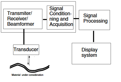
All ultrasound systems consist of five parts as shown in Fig.1 : the first includes an acoustical part, the transducer, which performs both the generation and the detection of ultrasound. This transducer may consist of one element (which has to move) or of multiple elements for multidimensional signal acquisition. The typical working frequency depends on the materials investigated by ultrasound. These frequencies are basically less than 500 KHz in air and range from 1 MHz in some liquids or biological tissue up to 50 or 100 MHz in some special applications.

Fig. 1. Ultrasound Image acquisition system
ONE DIMENSIONAL SIGNAL PROCESSING : TIME, FREQUENCY AND VELOCITY ESTIMATION
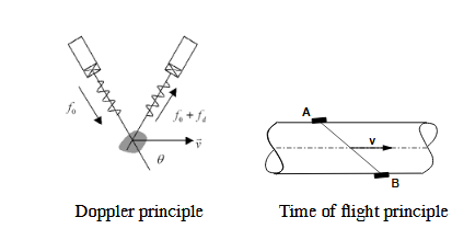
The primary aim of 1D US signal processing is to extract some parameters form the US signal for estimation or detection purpose. One important matter of concern is flow velocity estimation. Two kinds of techniques are used for this purpose.

Fig. 2. Velocity estimation principles
2D IMAGING
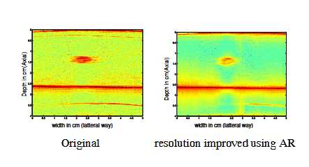
US Image processing has been mainly centered on 2D image manipulation. Basically, 2D US imaging systems operate in Brightness-mode or B-mode Fig.3. A typical B-mode image is obtained from of a set individual radio-frequency (RF) signals by filtering, envelope detection and then log- compression.

Fig. 3. B-mode image of resolution improvement
3D AND 4D IMAGING
Although 2D US imaging offers very acute understanding of the imaged media, its has some intrinsic limitations; the main of which being 2D viewing of 3D structures. This requires a mind reconstruction of actual 3D structures, which is in some situations difficult to achieve. A way to over- come this is to take multiple scans of some particular locations and to quantitatively analyze them afterwards. This is particularly time consuming. 3D and 4D may thus be used to overcome these limitations. 4D US imaging is real-time 3D US imaging in which time is taken as the fourth coordinate. The starting point of 3D/4D US imaging is, like in the 2D case, the acquisition system.
CONCLUSION
This paper has reviewed the main ultrasound imaging systems and techniques from 1D signals to 4D images. The part played by signal and image processing has become larger and larger, and current trends in new applications, both in industrial and biomedical fields, are a sign that this part will keep on growing. Finally, several practical developments have been presented and also discussed during the oral presentation in this special session.
Source: University of Tours
Authors: D. Kouame | J.M. Gregoire | L. Pourcelot | J.M Girault | M. Lethiecq and F. Ossant
>> 100+ Projects on Image Processing
>> 60+ Simple Biomedical Project Titles for Final Year Engineering Students
>> Matlab Projects for Biomedical Engineering Students
>> 50+ Matlab projects for Digital Image Processing for Final Year Students
>> More Matlab Projects on Signals and Systems for Final Year Students