ABSTRACT
For many health services in developing countries, patient identification is a fundamental need. In countries where no standard form of identification is available, this problem is exacerbated by a lack of literacy and also frequent errors in spelling and consistency. To address this need, we implemented two low-cost hand vein scanner devices for use with mobile devices.
The first scanner device employs the internal camera of the an Android smart phone along with a rechargeable infrared light (850nm) and an external optical filter; and the second scanner device employs a low-cost webcam, with integrated LEDs (940nm) and optical filter, which is powered directly from the Android tablet.
A single mobile app was developed for use with both scanner devices with the ability to adjust scanner settings, capture hand palm images, and annotate patient data. As an initial test of our scanner designs, we collected hand scans from 51 university students aged 18-34 using an IRB-approved protocol, and data was processed using a 2D-PCA biometric algorithm implemented on a PC using MATLAB software.
Using the standard FAR-FRR curve for biometric analysis, we were able to achieve an Equivalent Error Rate (EER) of 6.3% for the phone camera scanner, and 4.2% for the webcam scanner design. These results compare favorably with other published biometrics studies and demonstrate the potential of low-cost biometric devices that can be integrated with mobile phones and tablets.
CAPTURING THE HUMAN HAND VEIN PATTERN
Illumination and Optical Properties of the Human Hand
Proper illumination is needed in order to enhance the appearance of blood vessels in the hand. Some early vein detection methods employed far-infrared thermal imaging (FIR) in order to detect small temperature differences between the blood vessels and the surrounding tissues. However, newer lower-cost methods employ near-infrared (NIR) illumination and make use of the fact that near-infrared light is absorbed more strongly by human blood than the surrounding tissue, and thus appears darker.
Camera Properties
Standard hardware used for recording infrared or low-light images make use of charge-coupled device (CCD) cameras. However, the cameras found in modern consumer smart phones and webcams are manufactured using the standard silicon CMOS process which is less sensitive to infrared light. Furthermore, most consumer cameras, including smart phones, also contain an infrared filter blocking which is used to improve color rendition for photographs.
Image Processing and Data Analysis
Over the past decade, a wide variety of algorithms and image processing techniques have been published for vein biometrics, many of which have been adapted from hand palm biometric methods.
DEVICE IMPLEMENTATION

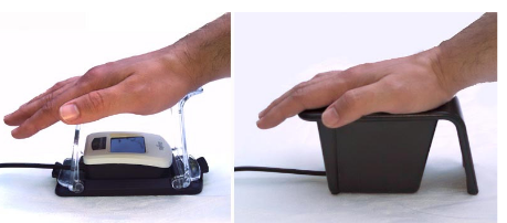
Fig 2. Two commercial vein scanning devices manufactured by Fujitsu
Comparing our conceptual model with two actual commercial vein scanning devices (Figure 2), we observed a practical need for a hand guide, to maintain a proper distance and orientation of the hand, and also an opaque shroud for the purpose of blocking external infrared light from lamps or sunlight.

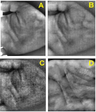
Fig. 3. (A, B, & C) Sample images from scanner 1 illuminated with 850nm IR along with ambient light leaking in through the edges
Fig. 3. (A, B, & C) Sample images from scanner 1 illuminated with 850nm IR along with ambient light leaking in through the edges: A= no filter, B=#87 filter (~795nm), C=#87C filter (~850nm). Comparing image A and B, one can see how the filter blocks reflected light from the skin surface and enhances the subsurface features. Image C shows increased graininess due to phone camera’s limited sensitivity at 850 nm. Image D is from scanner #2, illuminated with 940nm IR with #87C filter, showing increased sensitivity and greater level of detail.

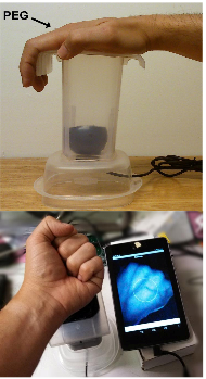
Fig. 5. (top) Side view of Scanner #2 prior to painting the inside black
Fig. 5. (top) Side view of Scanner #2 prior to painting the inside black. The placement of the hand and location of the webcam are clearly visible. (bottom) Demonstration of scanner showing live image of dorsal veins. The second scanner design is shown in Figure 5, and also consists of an opaque plastic lightbox. However the camera and the illumination were provided by an external webcam, described below.
EXPERIMENTAL STUDY

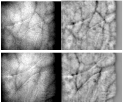
Fig. 6 Sample image data before (left) and after preprocessing (right)
A minimum amount of preprocessing was applied to each image, and consisted of a 0.5 pixel smoothing, 15-pixel high-pass filter, and a uniform contrast adjustment applied uniformly to all images, as shown in Figure 6. No vein contour extraction algorithm was used, and no correction was provided for misaligned or rotated hands.
RESULTS AND DISCUSSION
Data statistics
For each scanner, a total of 478 images were analyzed from 51 participants. Of these, 204 images (4 per subject) were used as training data, 51 images (1 per subject) were used as validation data, and 223 images (4 or 5 per subject) were used as test data.
False Accept Rate (FAR) vs False Reject Rate (FRR)
The results from both scanner devices are shown in Figure 5, with scanner 1 results plotted in red, and scanner 2 results plotted in blue. Both curves have a good basic square shape and approach the origin within a few percent. The point at which FAR=FRR, also known as the Equivalent Error Rate (EER) is 6.3% for scanner #1 and 4.2% for scanner #2, which compares favorably to other published vein biometric studies using using similar 2D-PCA algorithms.
Potential Applications and Threshold Setting
For those not familiar with biometric device statistics, it is worth noting that the exact operation point along the FAR- FRR curve depends on the threshold setting and the desired application. Sometimes articles or ads for biometric devices will provide an FAR or FRR statistic without providing EER or showing the complete FAR-FRR curve, which can be misleading.
CONCLUSIONS AND FUTURE WORK
We have successfully demonstrated the imaging capability of two biometric scanner designs based on mobile devices, with Equal Error Rates (EER) of 6.3% and 4.2%, respectively, which are within range of other biometric technologies and can be suitable for certain identification applications, or as a means of verification in places where identification infrastructure is lacking.
These results were also achieved using very small image sizes of 180 X 150 pixels, and 176 X 144 pixels, respectively, in order to demonstrate feasibility for implementation of the entire algorithm on a mobile phone. The two scanner device designs also have a very low materials cost (US$45, and US$15, respectively), with the second scanner design also eliminating the need for an external power source.
Through careful selection of the illumination frequency and optical filters, sufficient image quality was possible without the need for significant image enhancement. In addition, the mechanical design of the scanners helped minimize errors due to hand misalignment and obviated the need for extensive image processing to perform auto-correction.
The EER of these devices can perhaps be further improved through more advanced and efficient PCA algorithms without additional computational burden. This is currently being explored.
We hope that low-cost designs such as these, which minimize cost and algorithmic complexity will soon enable affordable and scalable biometric devices that can serve the needs of the global health community.
Source: IEEE
Authors: Richard Ribón Fletcher | Varsha Raghavan | Rujia Zha | Miriam Haverkamp | Patricia L. Hibberd
>> Matlab Projects for Biomedical Engineering Students
>> Image Processing Project Topics with Full Reports and Free Source Code
>> Huge List of Matlab Projects with Free Source Code
>> Matlab Projects Fingerprint Recognition and Face detection for Engineering Students
>> Medical Image Processing Projects using Matlab with Source Code for Engineering Students