ABSTRACT
At the moment, identification of blood disorders is through visual inspection of microscopic images of blood cells. From the identification of blood disorders, it can lead to classification of certain diseases related to blood. This paper describes a preliminary study of developing a detection of leukemia types using microscopic blood sample images.
Analyzing through images is very important as from images, diseases can be detected and diagnosed at earlier stage. From there, further actions like controlling, monitoring and prevention of diseases can be done. Images are used as they are cheap and do not require expensive testing and lab equipments. The system will focus on white blood cells disease, leukemia. The system will use features in microscopic images and examine changes on texture, geometry, color and statistical analysis.
Changes in these features will be used as a classifier input. A literature review has been done and Reinforcement Learning is proposed to classify types of leukemia. A little discussion about issues involved by researchers also has been prepared.
BACKGROUND
Blood is the main source of information that gives an indication of changes in health and development of specific diseases. Changes in the number or appearance of elements that formed will guide health condition of an individual.
RESEARCH METHODOLOGY
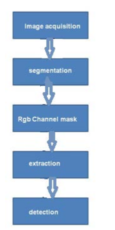
Research methodology that will be used in this research includes:
- Image Acquisition
- Pre-processing
- Segmentation
- Feature Extraction
- Classification

Figure: Implementation design process
DISCUSSION
Things that need to be discussed are to resolve some issues about the blood cells. One of the issue s is the problem on the blood cell itself. Claim that their system fail in classification processes for some of the blood cells. Some of the cells can be deformed to arbitrary shape due to environment pressure. Takes note on their algorithms that does not separate overlapping cells.
CONCLUSION
This research involves detecting the types of leukemia using microscopic blood sample images. The system will be built by using features in microscopic images by examining changes on texture, geometry, colors and statistical analysis as a classifier input. The system should be efficient, reliable, less processing time, smaller error, high accuracy, cheaper cost and must be robust towards varieties that exist in individual, sample collection protocols, time and etc. Information extracted from microscopic images of blood samples can benefit to people by predicting, solving and treating blood diseases immediately for a particular patient.
Source: JATIT
Authors: Fauziah Kasmin | Anton Satria Prabuwono | Azizi Abdullah
>> 60+ Simple Biomedical Project Titles for Final Year Engineering Students
>> Matlab Projects for Biomedical Engineering Students
>> Image Processing Project Topics with Full Reports and Free Source Code
>> 200+ Matlab Projects based on Control System for Final Year Students
